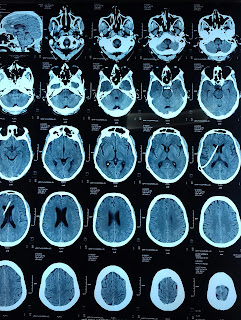
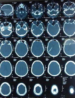
Ct scan of Brain: Ventriculoperitoneal shunt image
 |
| vp shunt ct scan |
A VP shunt, also known as a ventriculoperitoneal shunt, is a medical device commonly used to treat hydrocephalus, a condition characterized by the accumulation of excess cerebrospinal fluid (CSF) in the brain. The VP shunt provides a pathway for draining the excess fluid from the brain's ventricles to the peritoneal cavity in the abdomen, where it can be absorbed and eliminated.
Hydrocephalus occurs when there is an imbalance between the production and absorption of CSF, leading to increased pressure within the brain. This can result in various symptoms, including headaches, nausea, vomiting, visual disturbances, and cognitive impairment. If left untreated, hydrocephalus can cause permanent brain damage and potentially be life-threatening.
A VP shunt consists of several components:
1. Ventricular Catheter: This flexible tube is surgically placed into one of the brain's ventricles, typically the lateral ventricle. It allows for the drainage of excess CSF from the brain.
2. Valve System: The valve is a critical component of the VP shunt. It regulates the flow of CSF and helps maintain an appropriate pressure within the brain. The valve can have various designs, including fixed-pressure valves, adjustable-pressure valves, or programmable valves, which allow for customization based on the individual's needs.
3. Distal Catheter: This tube extends from the valve system and is tunneled under the skin, connecting the valve to the peritoneal cavity. It allows the CSF to be transported from the brain to the abdominal cavity, where it can be reabsorbed.
The VP shunt works by diverting the excess CSF from the brain to the peritoneal cavity, where it can be absorbed by the body. The valve system helps regulate the flow of CSF, preventing overdrainage or underdrainage. Regular monitoring and adjustment of the valve may be necessary to ensure optimal pressure within the brain.
The insertion of a VP shunt is typically performed under general anesthesia in a surgical setting. The surgeon makes small incisions in the scalp, creates a small hole in the skull (burr hole), and carefully places the ventricular catheter into the appropriate ventricle. The distal catheter is then tunneled under the skin and inserted into the peritoneal cavity. The valve system is connected to both catheters, facilitating CSF flow.
Following the surgery, patients may require a short hospital stay for observation. Pain medication and antibiotics may be prescribed to manage post-operative discomfort and reduce the risk of infection. Regular follow-up visits are essential to monitor the functioning of the VP shunt, check for any signs of infection or blockage, and make adjustments to the valve settings, if necessary.
While VP shunts are generally effective in managing hydrocephalus, there are potential risks and complications associated with the procedure. These include infection, blockage or malfunction of the shunt system, overdrainage or underdrainage of CSF, bleeding, and injury to surrounding structures. Prompt medical attention should be sought if any signs of shunt malfunction, such as headaches, changes in vision, or nausea, occur.
In conclusion, a VP shunt is a medical device used to treat hydrocephalus by providing a pathway for the drainage of excess CSF from the brain to the peritoneal cavity. It helps alleviate symptoms and manages the condition. Regular monitoring and follow-up care are crucial to ensure the proper functioning of the shunt and to address any complications that may arise.

